
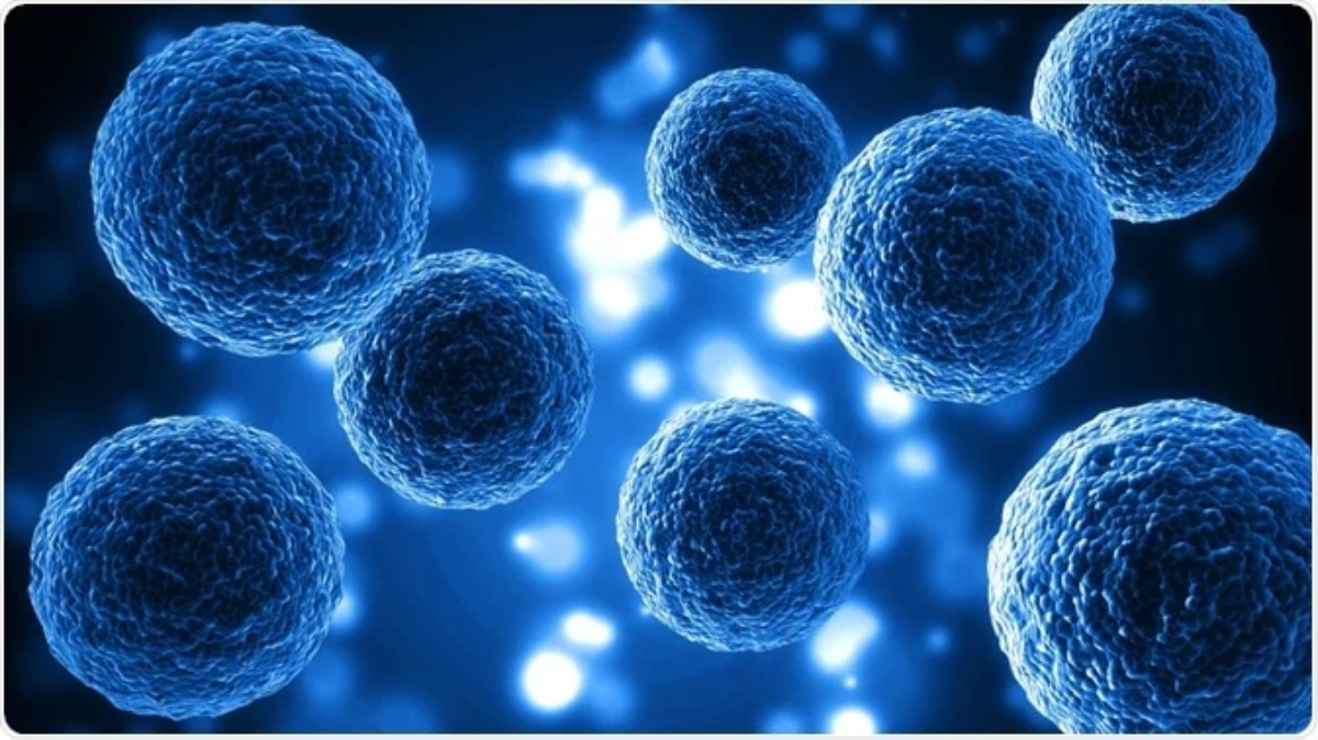
The Cell: Nature’s Microscopic Masterpiece – The Foundation of All Living Life
Beneath the limits of the naked eye lies one of nature’s most extraordinary creations: the cell. Known universally as the fundamental building block of every living organism, this tiny living unit functions as an independent, highly organized world. Even though cells vary dramatically in shape, size, and specialized roles, every living cell hosts thousands of organic molecules and sustains a continuous cascade of complex biochemical reactions. These reactions govern construction, breakdown, energy production, waste management, and communication – activities that collectively define the life of the entire organism, whose existence is simply the sum of all its cellular activities.
The journey toward discovering this microscopic wonder began in the 17th century. In 1665, English scientist Robert Hooke crafted a simple microscope and examined cork plant tissue. He observed regular, hollow compartments resembling honeycomb cells and coined the term “cell” – meaning a small cavity. Years later, in 1673, Antonie van Leeuwenhoek of the Netherlands built a more powerful microscope and became one of the first to observe animal cells. Nevertheless, the intricate details of cellular anatomy remained hidden for centuries; it was only with the advent of electron microscopes in the mid‑20th century that scientists finally gained the ability to map the cell’s inner machinery with precision.
Living organisms differ vastly in cellular composition. Single‑celled life such as bacteria relies on just one cell to perform every essential task: movement, respiration, metabolism, waste removal, and defense against threats. There are no specialized cell types here – one cell serves all purposes. By contrast, the human body is composed of approximately 50 to 100 trillion specialized cells. A single square inch of human skin contains roughly one million cells, while the brain holds more than thirty billion cells. Water makes up about 60 % of human body weight: 40 % resides inside cells, 15 % fills the extracellular space, and 5 % circulates within blood vessels. Cells absorb nutrients and oxygen from surrounding fluids and release metabolic wastes, which blood transports to the kidneys for elimination as urine.
Cells organize themselves into a beautifully layered hierarchy of life. Groups of similar cells form tissues; tissues combine to create organs; organs collaborate to form body systems. Nerve cells assemble into nervous tissue that builds the brain and spinal cord – the core of the nervous system. The esophagus, stomach, and intestines unite to create the digestive system. Each system carries out a distinct mission: the respiratory system captures oxygen and expels carbon dioxide; the digestive system breaks food down into simple, absorbable molecules. Even cell structure adapts to function: respiratory cells feature hair‑like cilia to filter and condition incoming air, while muscle cells are elongated to enable contraction and relaxation.
Biologically, cells fall into two primary groups defined by structural complexity. Prokaryotic cells, such as bacteria, lack a membrane‑bound nucleus; their genetic material floats freely within the cytoplasm, and they possess relatively few organelles. Eukaryotic cells, found in animals, plants, and humans, enclose their genetic material inside a distinct nucleus and contain a rich array of specialized organelles. A typical eukaryotic cell consists of three core components: the cell membrane, the cytoplasm with its organelles, and the nucleus.
The cell membrane acts as the cell’s intelligent boundary. Built mainly from phospholipids arranged in a double layer plus embedded proteins, it is semi‑permeable: it selectively controls the flow of nutrients, ions, hormones, and waste products, protects the cell, maintains its shape, recognizes foreign invaders, and facilitates communication with the external environment through receptors and gateways.
The cytoplasm is a dynamic, gel‑like matrix composed largely of water, proteins, carbohydrates, lipids, and minerals. It contains specialized structures called organelles, each assigned a unique role:
- Mitochondria: Known as the cell’s powerhouses, they generate energy; cells with high activity like muscle and sperm cells contain the most mitochondria.
- Golgi apparatus: Modifies, packages, and distributes proteins produced by ribosomes.
- Lysosomes: Function as cellular digestive systems, breaking down waste, damaged organelles, and invading particles.
- Endoplasmic reticulum: Divided into rough ER (protein synthesis) and smooth ER (lipid production, mineral metabolism, and hormone regulation).
- Ribosomes: Tiny structures that manufacture proteins for use inside or outside the cell.
- Centrioles, cilia, and flagella: Aid cell division, movement, and transport of materials across cell surfaces.
The nucleus serves as the cell’s information center. Enclosed by a double nuclear membrane with tiny pores, it contains nuclear sap, a nucleolus, and chromatin – DNA that carries all genetic instructions required to build proteins and guide cellular development.
One of life’s most fascinating phenomena is cell differentiation. Every cell in the human body originates from a single fertilized egg and inherits the same DNA. Yet chemical signals during embryonic development activate or silence specific genes. This determines whether a cell becomes a pancreatic beta‑cell producing insulin, a muscle cell packed with contractile filaments, or a nerve cell built for electrical signaling. This selective gene activity creates the incredible diversity of cell types that make up the human body.
This entire understanding rests upon the historic Cell Theory, developed by Schleiden and Schwann and later expanded by Rudolf Virchow. It states four fundamental truths: the cell is the basic structural unit of life; an organism’s activity arises from the combined actions of its cells; cellular biochemical functions depend on the coordinated work of organelles; and every cell comes only from a pre‑existing cell through division.
From Hooke’s first glimpse of cork cells to modern electron microscopy and molecular biology, the study of the cell has transformed medicine, biology, and our appreciation of life itself. This microscopic masterpiece proves that the most profound secrets of existence are often hidden within the smallest structures known to science.
Disclaimer: This article is popular‑science educational content only. It is intended for general knowledge and magazine reading and does not replace formal academic textbooks, laboratory training, or medical consultation.
Useful Links:
https://tech.nashs.online
https://nashs.online
https://fast-convert.net